Orthopedic and neurologic diseases are common in companion animals. In practice, it can sometimes be difficult to determine which system is abnormal in a given patient. There are important aspects of history acquisition, the examination, and recognizing specific conditions with pathognomonic findings that can help a practitioner distinguish between orthopedic and neurologic diseases.
The purpose of this article is to aid the general practitioner in determining if a patient is experiencing an orthopedic or a neurologic cause of dysfunction. Specifically, there are notable orthopedic conditions that may be considered "neurologic imposters." However, these orthopedic conditions lack key deficits consistent with neurologic dysfunction. These "neurologic imposters" are addressed below to aid the clinician in the evaluation of patients that are presented for gait dysfunction.
Using the neurologic exam to identify neurologic versus orthopedic disease
Figure 1 provides a summary chart of the segments of the spinal cord and expected neurologic findings.
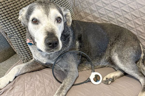
Figure 3: Doberman with left gastrocnemius rupture. Note the plantigrade stance and the curling of the digits, often confused with knuckling due to proprioceptive dysfunction. Courtesy Dr. Susan Arnold
Doberman with left gastrocnemius rupture.
Both pelvic limbs affected: Neurologically, conditions causing both pelvic limbs to be abnormal occur secondary either to a lesion in the spinal cord or in the peripheral nervous system. Regarding lesions in the spinal cord, issues with both the T3-L3 and L4-S1 regions of the spinal cord, will result in abnormalities in the pelvic limbs without concurrent abnormalities in the thoracic limbs.
These two localizations produce nearly opposing neurologic deficits. Patients with T3-L3 myelopathies have upper motor neuron paraparesis, characterized by stiffness and limited limb flexion, and general proprioceptive ataxia.
Patients with L4-S1 myelopathies have lower motor neuron paraparesis, characterized by weakness and difficulty bearing weight, with or without observable general proprioceptive ataxia. T3-L3 myelopathies result in increased pelvic limb tone and mild atrophy with time, whereas L4-S1 myelopathies result in decreased pelvic tone and often moderate to marked atrophy over a short period of time. Both can lead to postural reaction deficits. Patients with T3-L3 myelopathies have normal patellar and pelvic limb withdrawal reflexes, whereas patients with L4-S1 myelopathies have decreased patellar and pelvic limb withdrawal reflexes. Patients with lesions that span beyond L4-S1 to also include the S1-caudal regions of the spinal cord may also have absent perineal reflexes and lower motor neuron fecal and urinary incontinence.
Neurologic causes:
- Intervertebral disc extrusion or protrusion
- Lumbosacral stenosis or bilateral foraminal stenosis
- Degenerative myelopathy
- Severe kyphosis or other severe malformations
- Neoplasia (extradural masses affecting the vertebrae, intradural-extramedullary masses affecting the meninges, intramedullary masses)
- Discospondylitis with secondary empyema or vertebral column instability
- Meningomyelitis of unknown etiology
- Vertebral column trauma (fracture, subluxation, luxation)
- Fibrocartilaginous embolic myelopathy
- Geriatric onset laryngeal paralysis polyneuropathy (causing sciatic nerve dysfunction)
Neurologic imposters of bilateral pelvic limb dysfunction
Bilateral cranial cruciate ligament rupture: Cranial cruciate ligament rupture is the most common orthopedic condition managed in small animal practice, and generally only affects one limb at a time. In these typical cases, diagnosis is usually straightforward. However, cranial cruciate ligament rupture can affect both limbs simultaneously.
In these cases, clinicians may struggle to determine if the underlying cause of a patient's bilateral pelvic limb gait abnormality is orthopedic or neurologic. Patients with bilateral cranial cruciate ligament ruptures present with bilateral pelvic limb lameness. Careful observation will reveal dogs with bilateral cranial cruciate ligament rupture collapse on the stifle while weight-bearing. Performing drawer testing and cranial tibial thrust and observing stifle instability confirms the diagnosis.
Bilateral cranial cruciate ligament ruptures are sometimes confused for myelopathies because their bilateral instability can make them alter their gait to reduce their discomfort. However, patients with bilateral cranial cruciate ligament ruptures have no overt stiffness or weakness, no general proprioceptive ataxia, appropriate muscle tone, normal postural reactions, and normal spinal reflexes.
Dog with bilateral cranial cruciate ligament rupture.
Distal aortic thromboembolism: Most clinicians are able to identify distal aortic thromboembolic disease (saddle thrombus) in cats, which develop such emboli more frequently than dogs and with more consistent clinical signs. Distal aortic thromboemboli can cause more vague heterogeneous presentations in dogs. Most affected dogs have a chronic or insidious onset of signs over a period of weeks to months (Winter et al. 2012). Some dogs experience dramatic alterations in their pelvic limb dysfunction, oscillating from mild deficits to severe dysfunction in short periods of time.
Patients with distal aortic thromboemboli may mistakenly be considered to have neurologic dysfunction because loss of blood supply to their limbs results in weakness that appears very similar to lower motor neuron paraparesis. In severe cases, they may be paraplegic and also have reduced tone and spinal reflexes due to lack of arterial supply to their limbs. However, a critical observation in dogs with distal aortic thromboemboli is weak or absent femoral pulses. Therefore, any patient with pelvic limb dysfunction, especially if they have signs consistent with lower motor neuron dysfunction, should have femoral pulse quality assessed.
Dog with distal aortic thromboembolism.
All four limbs affected: Neurologically, in patients with all four limbs affected, there are three possible localizations provided they have normal mentation and a normal cranial nerve exam: C1-C5 myelopathy, C6-T2 myelopathy, and diffuse neuromuscular. Refer to Figure 1 for examination findings for patients with C1-C5 and C6-T2 myelopathies. Patients with diffuse neuromuscular disease have lower motor neuron tetraparesis, meaning they look weak, and either have exercise intolerance or take short, choppy steps when they walk. They also have difficulty bearing weight. They do not have general proprioceptive ataxia and also have normal postural reactions, and can have normal to absent limb reflexes, depending on the underlying etiology.
Neurologic causes:
- C1-C5 and C6-T2 myelopathies:
- Intervertebral disc extrusion or protrusion
- Cervical spondylomyelopathy
- Atlantoaxial subluxation or luxation
- Neoplasia (extradural masses affecting the vertebrae, intradural-extramedullary masses affecting the meninges, intramedullary masses)
- Meningomyelitis of unknown etiology
- Steroid-responsive meningitis arteritis (neck pain only, no deficits)
- Vertebral column trauma (fracture, subluxation, luxation)
- Fibrocartilaginous embolic myelopathy
- Diffuse neuromuscular disease:
- Polyradiculoneuritis
- Myasthenia gravis
- Tick paralysis
- Botulism
- Polymyositis
Neurologic imposter of dysfunction in all limbs
Immune-mediated polyarthritis: Immune-mediated polyarthritis is characterized by noninfectious inflammation of multiple joints. Dogs with this condition are reluctant to walk and exhibit a stiff, stilted and short, choppy gait in all limbs secondary to their discomfort. Some dogs also have accompanying neck pain.
Their abnormal gait can sometimes be confused for either C1-C5 myelopathy or diffuse neuromuscular disease. However, unlike patients with C1-C5 myelopathy, patients with immune-mediated polyarthritis have no general proprioceptive ataxia, and have normal postural reactions. Unlike patients with diffuse neuromuscular disease, patients with immune-mediated polyarthritis have normal spinal reflexes. Patients with immune-mediated polyarthritis often have an elevated core body temperature, palpable joint effusion, and evidence of pain with joint manipulation.
Conclusion
Neurologic dysfunction relative to a specific region of the nervous system results in consistent deficits across patients. Thus, recognizing the pattern of abnormalities expected with each localization is important in confirming that a patient presented for gait abnormality is truly neurologically abnormal.
Conversely, there are many musculoskeletal/orthopedic conditions that are rarely encountered and, therefore, may be challenging to recognize. Awareness of these uncommon conditions can aid the practitioner in diagnosis and treatment of patients with "neurologic imposter" conditions.
Susan A. Arnold, DVM, DACVIM (Neurology) is an assistant professor of neurology and neurosurgery at the University of Minnesota College of Veterinary Medicine. She is boarded within the American College of Veterinary Internal Medicine (Neurology). In her role, Dr. Arnold practices neurology and neurosurgery, trains interns and residents, and teaches veterinary medical students in both didactic and clinical settings.