A step-by-step photo guide to crown amputations

The author showcases how using less force and more finesse can help master these extractions

All photos by Dr. Jan Bellows

Introduction

Tooth extraction in veterinary patients is a delicate balance of skill, patience, and precision. Traditional extractions carry the risk of root fracture, hemorrhage, or even mandibular trauma—especially in patients with advanced periodontal disease. 1

Enter crown amputation-assisted extractions. This method prioritizes anatomical respect, surgical visibility, and patient safety while reducing procedural time and stress for the practitioner.

When to extract a tooth

Extractions are indicated in the following presentations:

  • Stage 4 periodontal disease
    Greater than 50 percent attachment loss, probing depths, Stage 3+ mobility, Stage 3 furcation, or gingival recession past the mucogingival junction.
  • Stage 3 periodontal disease
    Attachment loss of 25–50 percent. Recommended when home care is ineffective or not possible.
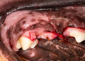
  • Fractured teeth
    Especially those with pulp exposure, internal resorption, where endodontic therapy isn't available, appropriate or accepted.
Crown root fracture
Figure 1: Complicated crown root fracture of the right maxillary fourth premolar. Photo courtesy Dr. Jan Bellows
  • Tooth resorption
    When advanced or communicating with the oral cavity requires extraction.
  • Feline gingivostomatitis
    Distal extractions for partial responders; full-mouth extractions for non-responders.
  • Supernumerary and persistent primary teeth
    Crowding and malocclusion risks call for timely removal. 

Complications of traditional extraction techniques

Even well-executed extractions can have setbacks. Complications include:

  • Root fracture
    Often from excessive force at the crown-root interface.
  • Hemorrhage
    Occurs when neurovascular bundles are inadvertently damaged while accessing alveolar bone.
  • Mandibular separation or fracture
    Especially in patients with periodontal bone loss and weakened jaw structure.

Crown amputation significantly reduces these risks by allowing for controlled, progressive removal of the tooth, starting at the crown. 

Why crown amputation?

Removing the crown first is a game-changer. It improves visibility, reduces leverage forces, and creates clean access to root structures. The result is fewer complications and better surgical outcomes. 2

Advantages include:

  • Smaller flap required
  • Less intraoperative bleeding
  • Easier and safer bone removal
  • Fewer root fractures
  • Shorter procedure times 

Instrumentation checklist

Include the following tools on your surgical tray:

  1. #15 or 15c Scalpel blade with handle – for gingival incisions
  2. #701 surgical bur – for moat creation and crown separation
  3. #2 flat molt elevator – flap elevation
  4. Wing-tipped elevators – for controlled root elevation, their unique shape allows for more efficient circumferential force application during elevation, minimizing torque stress on the bone.3
  5. Fine extraction forceps or root tip Pick – for root removal if needed
  6. Football diamond bur – smoothing alveolar bone
  7. 4-0 catgut or Monocryl with P-3 reverse cutting needle – suture closure

Operative technique: step-by-step

1. Local anesthesia

Administer appropriate regional blocks to ensure patient comfort and minimize bleeding.

2. Sulcular incision

Using a scalpel blade, incise the gingiva 360 degrees around the tooth, severing the coronal periodontal ligament.

3. Raise a buccal flap

Create a full thickness mucoperiosteal flap (envelope or pedicle) to expose the coronal buccal alveolus.

Separate the attached gingiva from the alveolar ridge
Figure 2: Molt elevator used to separate the attached gingiva from the alveolar ridge. 

4. Section the tooth into single rooted segments

Start sectioning at the furcation progressing coronally
Figure 3A: Start sectioning at the furcation progressing coronally.
Sectioning the palatal root
Fig 3B: Sectioning the palatal root. 


5. Horizontal crown sectioning

Section the crown horizontally at the CEJ.

Horizontal positioning of the bur for coronectomy
Figure 4: Horizontal positioning of the bur for coronectomy.

6. Create a moat or gutter at the cementoenamel junction (CEJ)

With a #701 surgical bur, circumferentially trough the supporting bone apical to the cementoenamel junction.

Creating the moat around each root
Figure 5: Creating the moat around each root.4

7. Wing-tipped elevator engagement

Apply moderate, controlled torsion circumferentially to mobilize the root.

 Inserting wing-tipped elevator between the sectioned roots
Figure 6: Inserting wing-tipped elevator between the sectioned roots.5

8. Extract root

Use a root tip pick or fine extraction forceps for final delivery.

Delivery of mesial root.
Figure 7A: Delivery of mesial root. Photos courtesy Dr. Jan Bellows
Delivery of distal root
Figure 7B: Delivery of distal root.
 Delivery of palatal root.
Figure 7C: Delivery of palatal root.

 9. Smooth bone margins

Use a football diamond bur to contour sharp bone ridges.

Alveolarplasty to smooth down the alveolar crest
Figure 8: Alveolarplasty to smooth down the alveolar crest.

 10. Flap closure

Enlarge flap if needed and suture using 4-0 catgut on a reverse cutting needle for tension-free primary closure.

Flap closed with 4-0 catgut
Figure. 9: Flap closed with 4-0 catgut 

Adding crown amputation: a win-win

This technique not only improves the clinical outcome but also:

  • Decreases surgical stress and time
  • Reduces postoperative complications
  • Enhances operator control
  • Shortens healing time

Veterinary dentistry continues to evolve, and with it, so should our surgical protocols. Adding crown amputation simplifies extractions while reducing risks and complications. By adopting this technique, you're not only preserving jaw integrity and ensuring complete removal, it's safer approaching roots with a "drone's eye" view. 8

Jan Bellows, DVM, DIPL, AVDC, ABVP, is one of six board-certified companion animal specialists (canine and feline) in Broward County, Florida. He is also one of 190 Board Certified Veterinary Dentists in the United States. He practiced in Pembroke Pines, Fla., for over twenty years before opening Hometown Animal Hospital and Dental Clinic in February 2000. That practice is both a Certified Specialty Facility and a Certified Hospital, certified by the American Animal Hospital Association. Dr. Bellows has received numerous awards, including the Florida Veterinary Medical Association Gold Star Award in 2012, and he has given numerous presentations on veterinary dentistry.

References

1. Wiggs, R. B., & Lobprise, H. B. 1997. Veterinary Dentistry: Principles and Practice. Philadelphia: Lippincott-Raven. pp. 302–305.
2. Niemiec, B. A. 2020. "Current Concepts in Surgical Tooth Extractions." Clinician's Brief, vol. 18, no. 4, pp. 47–51.
3. Reiter, A. M. 2008. "Small Animal Dental Equipment, Materials, and Techniques." In Verstraete, F. J. M., & Lommer, M. J. (Eds.), Oral and Maxillofacial Surgery in Dogs and Cats. Saunders Elsevier, pp. 10–25.
4. Bellows, J. 2019. "Techniques to Minimize Extraction Complications." Journal of Veterinary Dentistry, Vol. 36, No. 2, pp. 76–84.
5. Visser, E., & Tsugawa, A. J. 2021. "Risk Factors and Prevention of Root Fractures During Dental Extraction." Veterinary Clinics of North America: Small Animal Practice, Vol. 51, No. 3, pp. 529–543.
8. Verstraete, F. J. M., & Lommer, M. J. 2012. Oral and Maxillofacial Surgery in Dogs and Cats. 2nd ed., Saunders Elsevier, pp. 231–240.

Comments
Post a Comment

Comments